
Types of Skeletal Muscle Fibers
The mammalian body has three major types of skeletal muscle fibers: fast fibers, slow fibers, and intermediate fibers.
Fast Fibers
Most of the skeletal muscle fibers in the body are called fast fibers, because they can contract in 0.01 sec or less after stimulation. Fast fibers are large in diameter; they contain densely packed mofibrils, large glycogen reserves, and relatively few mitochondria. The tension produced by a muscle fiber is directly proportional to the number of sarcomeres, so muscles dominated by fast fibers produce powerful contractions. However, fast fibers fatigue rapidly because their contractions use ATP in massive amounts, so prolonged activity is supported primarily by anaerobic metabolism. Several other names are used to refer to these muscle fibers, including white muscle fibers, fast-twitch glycolytic fibers, and Type II-A fibers.
Slow Fibers
Slow fibers are only about half the diameter of fast fibers and take three times as long to contract after stimulation. Slow fibers are specialized to enable them to continue contracting for extended periods, long after a fast muscle would have become fatigued. The most important specializations improve mitochondrial performance. Slow muscle tissue contains a more extensive network of capillaries than is typical of fast muscle tissue and so has a dramatically higher oxygen supply. In addition, slow fibers contain the red pigment myoglobin (an oxygen-binding pigment that is especially common in slow skeletal muscle fibers and cardiac muscle cells). This globular protein is structurally related to hemoglobin, the oxygen-carrying pigment in blood. Both myoglobin and hemoglobin are red pigments that reversibly bind oxygen molecules. Although other muscle fiber types contain small amounts of myoglobin, it is most abundant in slow fibers. As a result, resting slow fibers contain substantial oxygen reserves that can be mobilized during a contraction. Because slow fibers have both an extensive capillary supply and a high concentration of myoglobin, skeletal muscles dominated by slow fibers are dark red. They are also known as red muscle fibers, slow-twitch oxidative fibers,and Type I fibers.
With oxygen reserves and a more efficient blood supply, the mitochondria of slow fibers can contribute more ATP during contraction. Thus, slow fibers are less dependent on anaerobic metabolism than are fast fibers. Some of the mitochondrial energy production involves the breakdown of stored lipids rather than glycogen, so glycogen reserves of slow fibers are smaller than those of fast fibers. Slow fibers also contain more mitochondria than do fast fibers.
Figure below compares the appearance of fast and slow fibers.
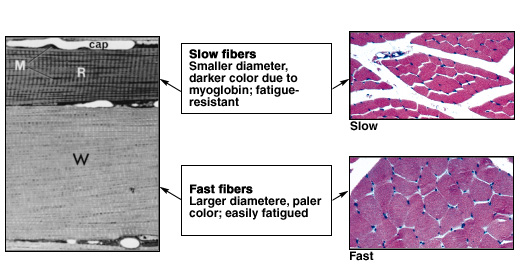
FIGURE – Fast versus Slow Fibers. (a)The slender, slow fiber (R for red) has more mitochondria (M) and a more extensive capillary supply (cap) than does the fast fiber (W for white). (LM X 783) (b) Notice the difference in the sizes of slow fibers, above, and of fast fibers, below. (LM X 171)
Intermediate Fibers
The properties of intermediate fibers are intermediate between those of fast fibers and slow fibers. In appearance, intermediate fibers most closely resemble fast fibers, for they contain little myoglobin and are relatively pale. They have a more extensive capillary network around them, however, and are more resistant to fatigue than are fast fibers. Intermediate fibers are also known asfast-twitch oxidative fibers and Type II-B fibers.
The three types of muscle fibers are compared in Table below. In muscles that contain a mixture of fast and intermediate fibers, the proportion can change with physical conditioning. For example, if a muscle is used repeatedly for endurance events, some of the fast fibers will develop the appearance and functional capabilities of intermediate fibers. The muscle as a whole will thus become more resistant to fatigue.
Properties of Skeleteal Muscle Fiber Types |
|||
Property |
Slow | Intermediate | Fast |
Crosss-sectional diameter |
Small | Intermediate | Large |
Tension |
Low | Intermediate | High |
Contraction speed |
Slow | Fast | Fast |
Fatigue resistance |
High | Intermediate | Low |
Color |
Red | White | White |
Myoglobin content |
High | Low | Low |
Capillary supply |
Dense | Intermediate | Scarce |
Mitochodria |
Many | Intermediate | Few |
| Glycolitic-enzime concentration in sarcoplasm | Low | High | High |
| Substrates used for ATP generation during contraction | Lipids, carbohydrates, amino acids (aerobic) | Primarily carbohydrates (anaerobic) | Carbohydrates (anaerobic) |
Alternative names |
Type I, S (slow), red, SO (slow oxidizing), slow-twitch oxidative | Type II-B, FR (fast resistant), fast-twitch oxidative | Type II-A, FF (fast fatigue) white, fast-twitch glycolitic |
Muscle Performance and the Distribution of Muscle Fibers
The percentages of fast, intermediate, and slow fibers in a skeletal muscle can be quite variable. Muscles dominated by fast fibers appear pale and are often called white muscles. Chicken breasts contain “white meat” because chickens use their wings only for brief intervals, as when fleeing from a predator, and the power for flight comes from fast fibers in their breast muscles. As we learned earlier, the extensive blood vessels and myoglobin in slow fibers give these fibers a reddish color; muscles dominated by slow fibers are therefore known as red muscles. Chickens walk around all day, and the movements are performed by the slow fibers in the “dark meat” of their legs.
Most mammalian muscles contain a mixture of fiber types and so appear pink. However, there are no slow fibers in muscles of the eye or hand, where swift but brief contractions are required. Many back and calf muscles are dominated by slow fibers; these muscles contract almost continuously to maintain an upright posture. The percentage of fast versus slow fibers in each muscle is genetically determined. As we noted earlier, the proportion of intermediate fibers to fast fibers can increase as a result of athletic training.
Muscle Hypertrophy
As a result of repeated, exhaustive stimulation, muscle fibers develop more mitochondria, a higher concentration of glycolytic enzymes, and larger glycogen reserves. Such muscle fibers have more myofibrils than do muscles that are less used, and each myofibril contains more thick and thin filaments. The net effect is hypertrophy, (an increase in the size of tissue without cell division) or an enlargement of the stimulated muscle. The number of muscle fibers does not change significantly, but the muscle as a whole enlarges because each muscle fiber increases in diameter. Hypertrophy occurs in muscles that have been repeatedly stimulated to produce near-maximal tension. The intracellular changes that occur increase the amount of tension produced when these muscles contract. A champion weight lifter or bodybuilder is an excellent example of hypertrophied muscular development.
Physical Conditioning
Physical conditioning and training schedules enable athletes to improve both power and endurance. In practice, the training schedule varies depending on whether the activity is supported primarily by aerobic or anaerobic energy production.
Anaerobic endurance is the length of time muscular contraction can continue to be supported by glycolysis and by the existing energy reserves of ATP and CP. Anaerobic endurance is limited by
(1) the amount of ATP and CP on hand,
(2) the amount of glycogen available for breakdown, and
(3) the ability of the muscle to tolerate the lactic acid generated during the anaerobic period.
Typically, the onset of muscle fatigue occurs within 2 minutes of the start of maximal activity. Activities that require above-average levels of anaerobic endurance include a 50-meter dash or swim, a pole vault, and a weight-lifting competition. These activities involve the contractions of fast fibers. The energy for the first 10–20 seconds of activity comes from the ATP and CP reserves of the cytoplasm. As these reserves dwindle, glycogen breakdown and glycolysis provide additional energy. Athletes training to improve anaerobic endurance perform frequent, brief, intensive workouts that stimulate muscle hypertrophy.
Aerobic endurance is the length of time a muscle can continue to contract while supported by mitochondrial activities. Aerobic endurance is determined primarily by the availability of substrates for aerobic respiration, which the muscle fibers can obtain by breaking down carbohydrates, lipids, or amino acids. Initially, many of the nutrients catabolized by the muscle fiber are obtained from reserves in the sarcoplasm. Prolonged aerobic activity, however, must be supported by nutrients provided by the circulating blood. During exercise, blood vessels in the skeletal muscles dilate, increasing blood flow and thus bringing oxygen and nutrients to the active muscle tissue. Warm-up periods are therefore important not only in that they take advantage of treppe but also because they stimulate circulation in the muscles before the serious workout begins. Because glucose is a preferred energy source, aerobic athletes such as marathon runners typically “load” or “bulk up” on carbohydrates for the last three days before an event. They may also consume glucose-rich “sports drinks” during a competition
Training to improve aerobic endurance generally involves sustained low levels of muscular activity. Examples include jogging, distance swimming, and other exercises that do not require peak tension production. Improvements in aerobic endurance result from altering the characteristics of muscle fibers and improving the performance of the cardiovascular system:
- Altering the characteristics of muscle fibers.
The composition of fast and slow fibers in each muscle is genetically determined, and individual differences are significant. These variations affect aerobic endurance, because a person with more slow fibers in a particular muscle will be better able to perform under aerobic conditions than will a person with fewer. However, skeletal muscle cells respond to changes in the pattern of neural stimulation. Fast fibers trained for aerobic competition develop the characteristics of intermediate fibers, and this change improves aerobic endurance.
- Improving cardiovascular performance.
Cardiovascular activity affects muscular performance by delivering oxygen and nutrients to active muscles. Physical training alters cardiovascular function by accelerating blood flow, thus improving oxygen and nutrient availability.
Aerobic activities do not promote muscle hypertrophy. Many athletes train using a combination of aerobic and anaerobic exercises so that their muscles will enlarge and both anaerobic and aerobic endurance will improve. These athletes alternate an aerobic activity, such as swimming, with sprinting or weight lifting. The combination is known as interval training or cross-training.Interval training is particularly useful for persons engaged in racquet sports, such as tennis or squash, which are dominated by aerobic activities but are punctuated by brief periods of anaerobic effort.